

Thinking as a Hobby
3478463 Curiosities served
Scientific Imagery
Previous Entry :: Next Entry
Read/Post Comments (0)
Mindblog links to an interesting article on the topic of how to render scientific information graphically, specifically scientific imagery related to biology.
It's especially interesting to me since I've been focused a lot on the cortical minicolumn, reading many papers on cortical anatomy, and when you come across a rendering of a minicolumn, it almost always looks something like this:
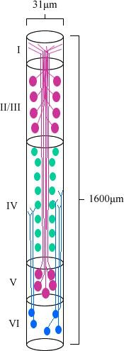
This is my own adaptation, from a drawing in:
Jones E.G. (2000) "Microcolumns in the Cerebral Cortex." Procedures of the National Academy of Sciences, 97: 5019–5021.
This is a rendering of a cortical minicolumn from a macaque monkey, and one of the things I didn't notice, and the authors never seem to draw attention to, is that the image is not drawn to scale. That is, the transverse diameter of the cylinder containing the minicolumn is about 30 micrometers, and the height traverses the entire width of the cortex, which is typically 2-4 millimeters, or 2,000-4,000 micrometers (in this case, 1,600 micrometers).
In fact, I've never seen one drawn to scale, so I did one myself:
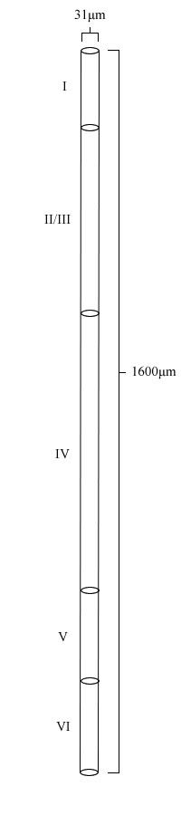
I understand why they skewed the minicolumn for visual purposes, but I think if they do that, they should draw attention to it in the text, or in the figure caption. In this case, I'm not even sure it was a good idea. I don't think the to-scale version, even with the cells and dendrites added, would be that bad to look at.
It's especially interesting to me since I've been focused a lot on the cortical minicolumn, reading many papers on cortical anatomy, and when you come across a rendering of a minicolumn, it almost always looks something like this:
This is my own adaptation, from a drawing in:
Jones E.G. (2000) "Microcolumns in the Cerebral Cortex." Procedures of the National Academy of Sciences, 97: 5019–5021.
This is a rendering of a cortical minicolumn from a macaque monkey, and one of the things I didn't notice, and the authors never seem to draw attention to, is that the image is not drawn to scale. That is, the transverse diameter of the cylinder containing the minicolumn is about 30 micrometers, and the height traverses the entire width of the cortex, which is typically 2-4 millimeters, or 2,000-4,000 micrometers (in this case, 1,600 micrometers).
In fact, I've never seen one drawn to scale, so I did one myself:
I understand why they skewed the minicolumn for visual purposes, but I think if they do that, they should draw attention to it in the text, or in the figure caption. In this case, I'm not even sure it was a good idea. I don't think the to-scale version, even with the cells and dendrites added, would be that bad to look at.
Read/Post Comments (0)
Previous Entry :: Next Entry
Back to Top

